CaNO: The Quiet Signal from the Deep Lungs
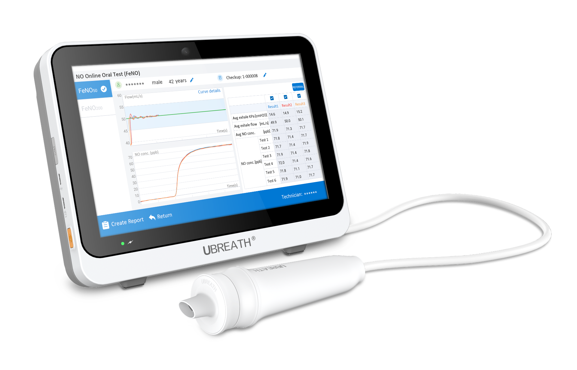
When you breathe out, you exhale more than just carbon dioxide and water vapor. Your breath carries trace amounts of nitric oxide (NO), a gas that serves as a biological messenger. For decades, doctors have used a simple breath test called FeNO (fractional exhaled nitric oxide) to detect inflammation in the large airways—the bronchial tubes that carry air into and out of the lungs. But FeNO tells only part of the story. The deepest regions of the lungs, where oxygen actually enters the bloodstream, have their own story to tell. That story is written in a lesser-known signal called CaNO.
What Exactly Is CaNO?
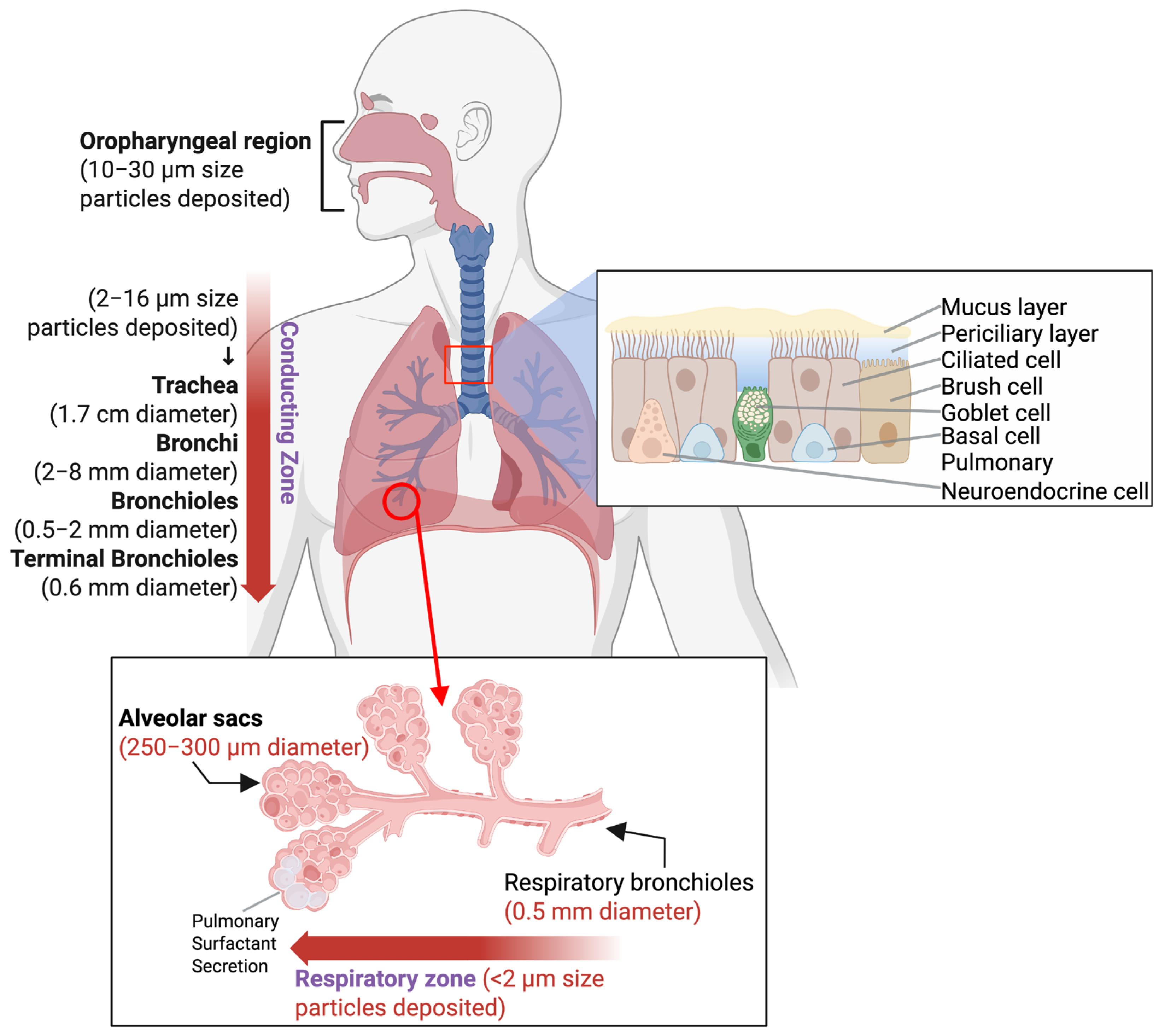
CaNO stands for alveolar concentration of nitric oxide—the amount of NO gas originating from the alveoli, the tiny air sacs at the very end of the respiratory tree where gas exchange takes place. If FeNO is a window into the bronchial tubes, CaNO is a window into the lung's deepest parenchyma.
The lungs can be thought of as a two-compartment system. The first compartment is the conducting airways—the bronchi and bronchioles—which produce NO mainly through the activity of inducible nitric oxide synthase (iNOS) in inflamed epithelial cells. The second compartment is the alveolar region, where NO behaves very differently. Because NO binds avidly to hemoglobin, alveolar NO is normally present at extremely low concentrations. When CaNO rises, it signals that something is happening in the alveolar tissue itself: inflammation, oxidative stress, or disruption of the delicate alveolar-capillary barrier.
How Is CaNO Measured?
Measuring CaNO is not as simple as blowing into a handheld device. Because FeNO depends heavily on how fast you exhale, clinicians use a technique called multiple-flow exhaled NO analysis. The subject exhales at several different flow rates—typically 10, 30, 100, and 200 mL/s—and a mathematical model derived from the two-compartment theory calculates CaNO by separating the alveolar contribution from the bronchial contribution.
The 2011 European Respiratory Society technical standards formally proposed this multi-parameter approach, which yields not only CaNO but also CawNO (bronchial wall NO concentration), JawNO (bronchial NO flux), and DawNO (bronchial NO diffusivity). This matters because FeNO alone can be misleading. In critically ill patients with excessive mucus secretion, for instance, the diffusion barrier thickens, DawNO drops, and FeNO may appear falsely low even when airway inflammation is severe. CaNO bypasses this problem by focusing on the alveolar compartment directly.
In healthy adults, the average CaNO is approximately 3.04 ppb (parts per billion), though reference values vary with age and smoking status. Smokers tend to have lower CaNO than non-smokers, likely because chronic smoke exposure alters alveolar NO dynamics.
Why CaNO Matters in Asthma
Asthma has traditionally been viewed as a disease of the large airways, and FeNO has become a standard tool for monitoring eosinophilic airway inflammation. But a growing body of evidence shows that asthma also affects the small airways—bronchioles less than 2 mm in diameter—and that this peripheral involvement is often missed by conventional FeNO testing.
CaNO fills this gap. In patients with severe or treatment-resistant asthma, CaNO levels are significantly higher than in healthy individuals or those with mild asthma, even when bronchial NO parameters look relatively normal. A landmark study found that the alveolar component of exhaled NO is specifically associated with lack of asthma control in patients with mild, untreated asthma. In other words, a patient might have a reassuring FeNO reading yet still have poorly controlled disease driven by inflammation deep in the small airways—a signal that only CaNO can detect.
This has practical implications. CaNO has been used to diagnose small airway dysfunction in stable asthma patients, with an optimal cutoff value of 5.3 ppb. In children with cough-variant asthma, CaNO has shown diagnostic value that may even exceed that of FeNO₅₀, the conventional measure of large-airway NO. For clinicians managing asthma, CaNO offers a more complete picture: it tells them not just whether the bronchi are inflamed, but whether the disease has reached the quiet, deep regions of the lung where conventional treatments like inhaled corticosteroids may not deposit effectively.
CaNO in COPD and Interstitial Lung Disease
Chronic obstructive pulmonary disease (COPD) is characterized by persistent airflow limitation, but its earliest pathological changes begin in the small airways, long before symptoms like breathlessness become apparent. CaNO has emerged as a promising biomarker for this peripheral airway inflammation. Studies comparing FeNO₂₀₀ and calculated CaNO in COPD patients suggest that CaNO may be a better indicator of distal small airway inflammation, which is the risk factor for COPD development.
The picture is even more striking in interstitial lung diseases (ILDs), a heterogeneous group of over 200 conditions that scar and stiffen the lung tissue. Because ILD primarily affects the alveolar interstitium and alveolar epithelium, CaNO is a far more relevant biomarker than FeNO. A systematic review concluded that CaNO is a promising marker of parenchymal inflammation in ILDs, reflecting tissue damage and altered gas diffusion. In systemic sclerosis–associated ILD, CaNO correlates with the severity of lung involvement as assessed by high-resolution CT and may even detect early lung involvement before radiological changes appear. Perhaps most importantly, in idiopathic pulmonary fibrosis—the most severe form of ILD—higher CaNO levels are associated with worse survival, suggesting that CaNO could serve as a prognostic biomarker to guide clinical decision-making.
The Road Ahead
CaNO is not yet a routine clinical test in most respiratory clinics, and formal trials are still needed to establish its added value over conventional FeNO in guiding treatment. Several challenges remain: measurement protocols need further standardization, reference values across different age groups and ethnic populations are still being refined, and the cost of multi-flow NO analyzers limits widespread adoption.
But the scientific rationale for CaNO is compelling. The lungs are not a single uniform organ; inflammation in the large airways and inflammation in the alveolar tissue are distinct processes with different clinical meanings. FeNO has given us a powerful tool for the former. CaNO extends that power to the latter—to the quiet, deep regions where the true work of breathing happens. As respiratory medicine moves toward increasingly personalized approaches, CaNO offers a non-invasive, repeatable way to listen to what the alveoli are saying. And sometimes, the quietest signals carry the most important messages.