Chronic Airway Diseases: When Breathing Becomes a Long-Term Challenge—and How Doctors Tell Them Apart
Breathing feels effortless until it isn’t. For hundreds of millions of people worldwide, the simple act of moving air in and out of the lungs is a daily struggle caused by a group of conditions known as chronic airway diseases. These are long-term conditions that affect the tubes—the airways—that carry air into and out of the lungs. They cannot usually be cured, but they can be managed, and the first step toward management is an accurate diagnosis.
Three of the most common chronic airway diseases are asthma, chronic obstructive pulmonary disease (COPD), and bronchiectasis. They share overlapping symptoms—cough, breathlessness, wheeze, and sputum production—but their underlying biology and their diagnostic requirements are not the same. Understanding how doctors distinguish among them is essential, because the right diagnosis leads to the right treatment.
A Scale That Is Hard to Ignore
The burden of these diseases is enormous. COPD alone affects approximately 480 million people globally and causes about 3.7 million deaths each year, making it the third leading cause of death worldwide. Asthma affected an estimated 363 million people in 2023 and caused 442,000 deaths. Bronchiectasis, once considered rare, is now recognized as far more common than previously thought, with reported prevalence ranging from 52 to over 1,400 cases per 100,000 adults, depending on the region and the population studied. These are not marginal conditions. They are among the most common chronic diseases of the respiratory system.
Asthma: Variable and Reversible
Asthma is best understood as a condition of variable airflow limitation. The airways are inflamed and hyperresponsive, so they tend to narrow in episodes—sometimes triggered by allergens, exercise, cold air, or infections—and then widen again. The hallmark symptom pattern is variation: symptoms come and go, and they vary in intensity over time.
The diagnostic key follows directly from that definition. Doctors look for evidence that airflow limitation is variable. The most common way to do this is spirometry with a bronchodilator reversibility test—lung function measured before and after inhaling a short-acting bronchodilator such as salbutamol. A positive response, consistent with asthma, is an increase in FEV₁ (forced expiratory volume in one second) of at least 12% and at least 200 mL from baseline. When spirometry is not available or results are normal but symptoms are typical, other tools can support the diagnosis, including peak expiratory flow variability, elevated fractional exhaled nitric oxide (FeNO), or elevated blood eosinophils. FeNO and eosinophils are markers of Type 2 inflammation, which underlies many cases of asthma, but they are supportive rather than definitive: elevated levels can occur in other conditions, and normal levels do not rule asthma out.
COPD: Persistent and Not Fully Reversible
COPD is defined by persistent airflow obstruction that is not fully reversible. It typically arises after long-term exposure to inhaled irritants—most commonly cigarette smoke, but also biomass smoke, occupational dusts, and air pollution. The symptoms are chronic and progressive: breathlessness on exertion, chronic cough, and sputum production.
The diagnostic standard for COPD is a post-bronchodilator FEV₁/FVC ratio below 0.70. This fixed threshold, recommended by the Global Initiative for Chronic Obstructive Lung Disease (GOLD), confirms that airflow obstruction persists even after the airways have been given the opportunity to relax with a bronchodilator. Spirometry is essential here: without it, COPD can be both overdiagnosed and underdiagnosed. GOLD has emphasized that many patients remain undiagnosed, and that making the interpretation of spirometry simpler and more consistent is a priority for improving case finding. Pre-bronchodilator spirometry can help rule COPD out, but post-bronchodilator measurement is required to confirm it, because some patients show a meaningful change in lung volumes only after bronchodilation.
Bronchiectasis: Structural Damage You Can See
Bronchiectasis is different in kind. It is not primarily defined by a pattern of airflow limitation on spirometry but by permanent, abnormal widening of the airways caused by chronic inflammation and infection. The airways lose their normal tapering and become flabby, scarred, and prone to pooling mucus. The classic clinical picture is a persistent cough producing copious sputum, often with recurrent respiratory infections, and sometimes with hemoptysis (coughing up blood).
The diagnostic gold standard is high-resolution computed tomography (HRCT) of the chest. The radiological criteria include an increased bronchoarterial ratio (the airway lumen is larger than its adjacent artery), lack of normal airway tapering, and visibility of airways within 1 cm of the pleural surface—signs that the airways are dilated and extend farther out into the lung than they should. HRCT is not merely confirmatory; it is the test that establishes the diagnosis. This matters because bronchiectasis can coexist with asthma or COPD, and it may be missed if clinicians assume that a patient’s chronic cough and sputum are fully explained by the other conditions.
Why Diagnosis Is Not Always Straightforward
If the distinctions were clean, diagnosis would be simple. In reality, the boundaries are blurred. A substantial proportion of patients—perhaps around 30% of those seen for airway disease—have overlapping features or mixed inflammatory profiles. Asthma, COPD, and bronchiectasis can coexist; a patient may have COPD as the dominant process but also meet criteria for bronchiectasis on imaging, or have asthma with a component of fixed airflow obstruction. Bronchodilator responsiveness, once thought to neatly separate asthma from COPD, can be observed in all three conditions. Symptoms alone—cough, breathlessness, wheeze—are not specific enough to reliably distinguish among them.
This overlap has real consequences. The rise of treatments targeted at specific disease mechanisms, such as biologic therapies for severe asthma or therapies specifically studied in bronchiectasis, makes diagnostic precision more important than ever. A drug that reduces exacerbations in bronchiectasis may not be appropriate for COPD, and a biologic that transforms the life of a patient with severe eosinophilic asthma is not interchangeable with standard COPD therapy. Accurate diagnosis is no longer an academic exercise; it determines whether a patient receives a treatment that can actually change the course of their disease.
The Diagnostic Toolbox, in Brief
Clinicians approach chronic airway disease diagnosis with a combination of tools, each answering a different question.
Spirometry measures how much air a person can forcefully exhale and how quickly. It is the central functional test. A reduced FEV₁/FVC ratio indicates airflow obstruction; the change after a bronchodilator helps distinguish variable from persistent obstruction. Spirometry requires patient effort and coaching, and in primary care settings it can be technically challenging, which is one reason underdiagnosis persists.
HRCT provides structural information. It is essential for diagnosing bronchiectasis and can also reveal emphysema, airway wall thickening, mucus plugging, and other features that support or complicate a diagnosis of COPD or asthma.
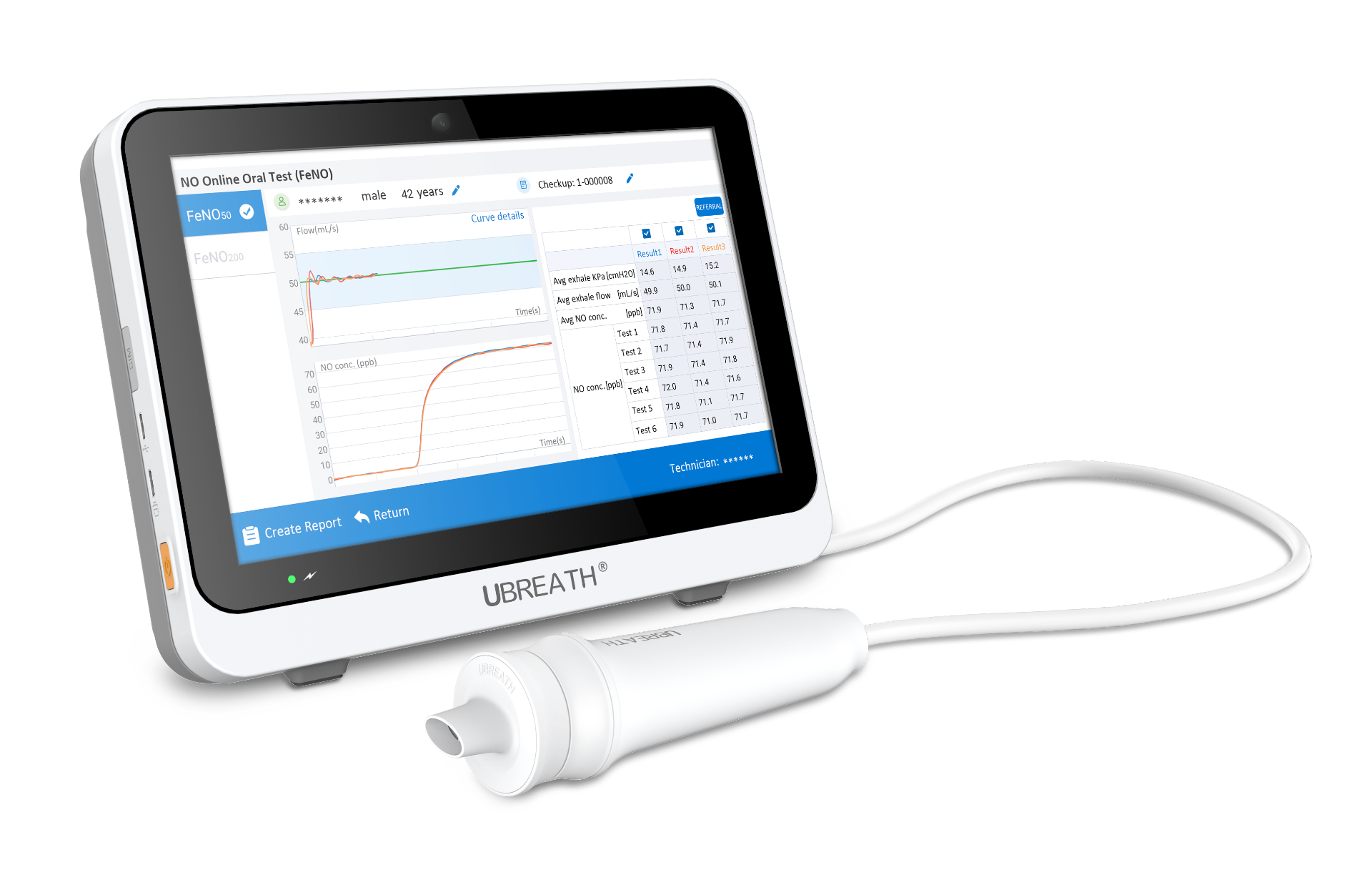
Biomarkers such as FeNO and blood eosinophils offer a window into the inflammatory profile. They are most useful in asthma, where elevated Type 2 markers support the diagnosis and can guide the use of inhaled corticosteroids and biologic therapies. They are not standalone diagnostic tests, but they add a dimension that spirometry and imaging cannot provide.
Clinical history remains indispensable. The pattern of symptoms over time—whether they are episodic and variable, chronic and progressive, or dominated by high-volume sputum—shapes the pre-test probability and determines which tests are most likely to be informative.
The Bottom Line
Chronic airway diseases are common, burdensome, and often underrecognized. Asthma, COPD, and bronchiectasis are not mutually exclusive categories; they exist on a landscape of overlapping symptoms and shared risk factors, with distinct but sometimes intersecting mechanisms. The diagnostic process is not a single test but a structured inquiry: confirming airflow obstruction with spirometry when the pattern suggests asthma or COPD, visualizing the airways with HRCT when bronchiectasis is suspected, and using biomarkers and clinical history to refine the picture. For patients, the message is that chronic breathlessness, cough, or sputum production deserves a proper evaluation—not because a label is inherently valuable, but because the right diagnosis opens the door to the right treatment.