FnNO: The Nasal Window into Upper Airway Health
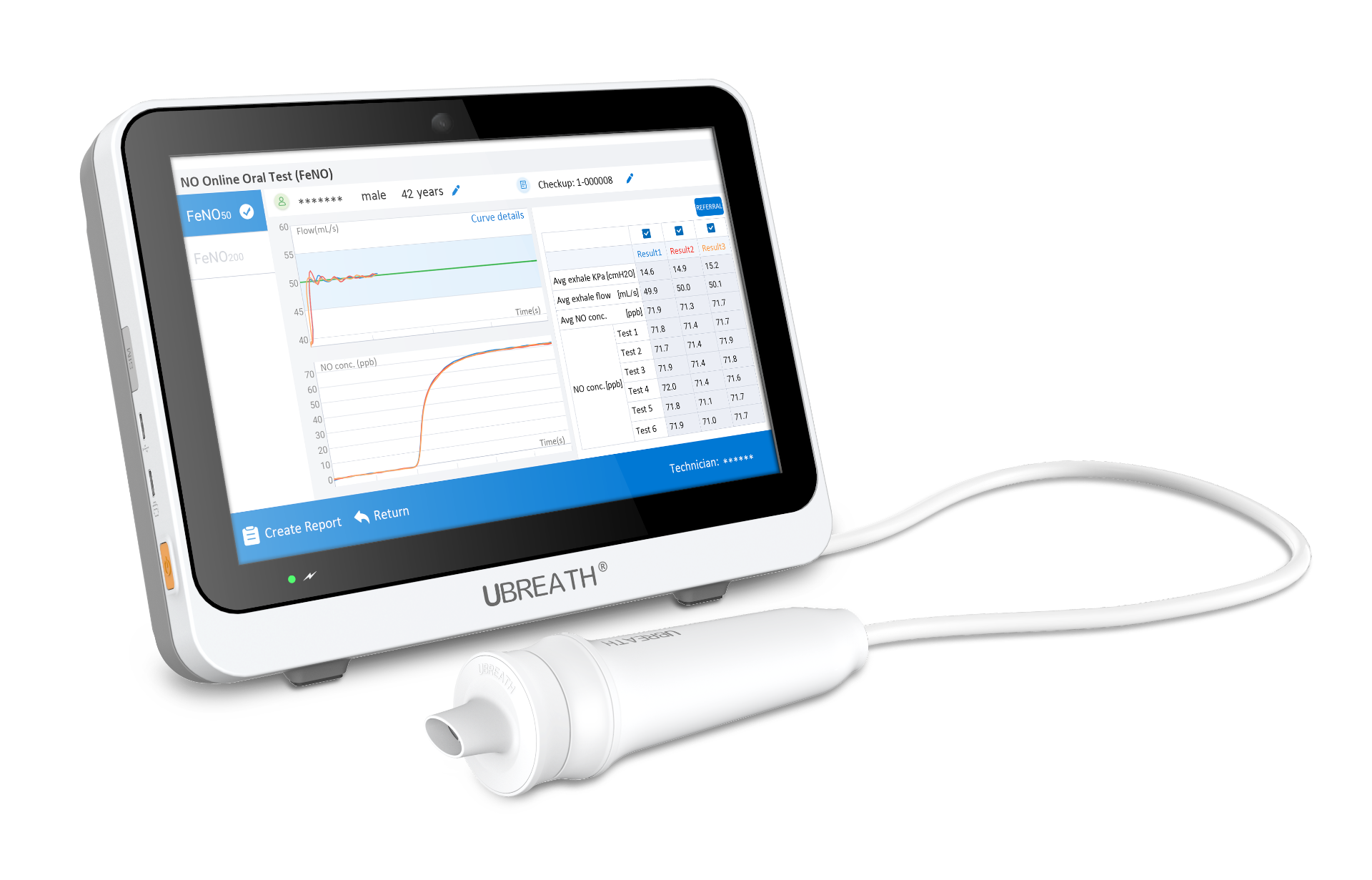
When we think about lung function testing, we usually picture spirometry—forceful blows into a tube that measure how much and how fast air moves. But there is another, quieter test that looks at a different part of the airway: fractional nasally exhaled nitric oxide, or FnNO. It measures nitric oxide (NO) in air exhaled through the nose, and it offers a non-invasive glimpse into inflammation in the nasal cavity and sinuses—the upper airway that is often overlooked in respiratory assessments.
What Exactly Is FnNO?
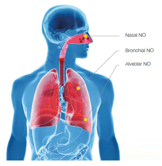
Nitric oxide is a gas produced by cells lining the respiratory tract. In the lower airways, NO is largely generated by inducible nitric oxide synthase (iNOS) in response to type 2 inflammation, which is why exhaled NO through the mouth (FeNO) has become a familiar biomarker in asthma. The nose is different. The paranasal sinuses constitutively produce large amounts of NO through endothelial nitric oxide synthase (eNOS), and this NO flows out through the nasal passages. When we measure FnNO, we are sampling that sinus-derived NO, and the value reflects the status of the nasal and sinus mucosa.
In healthy individuals, FnNO is typically in the range of 250 to 500 parts per billion (ppb) . The default sampling flow for FnNO is 10 mL/s, and the result is written as FnNO₁₀ . Because NO production and transport depend on sinus ostia patency, mucosal swelling, and ciliary function, FnNO can move in either direction—up or down—depending on what is happening in the upper airway.
Why the Upper Airway Deserves Its Own Biomarker
The “united airway” concept recognizes that the nose and lungs are not separate compartments. Allergic rhinitis frequently coexists with asthma, and poorly controlled upper airway inflammation can worsen lower airway symptoms. Yet standard pulmonary function tests assess only the lower airway. FnNO fills a gap: it provides an objective, point-of-care measure of upper airway inflammatory activity.
This matters clinically because FnNO behaves in distinct ways across different nasal conditions. In allergic rhinitis, FnNO is elevated, and the degree of elevation correlates with disease severity—the more active the allergic inflammation, the higher the nasal NO. In chronic rhinosinusitis, the picture is more nuanced. When sinus ostia are blocked by swollen mucosa, NO produced inside the sinuses cannot escape, and FnNO falls below normal. If eosinophilic inflammation coexists, FnNO may instead rise, so interpretation requires attention to ostial patency and the underlying inflammatory phenotype.
FnNO in Primary Ciliary Dyskinesia: A Screening Test with Real Power
The most established clinical role for FnNO is in the diagnostic pathway for primary ciliary dyskinesia (PCD), a rare genetic disorder in which impaired ciliary motion leads to chronic sinopulmonary infection, bronchiectasis, and—in about half of cases—situs inversus. PCD is often diagnosed late, and definitive testing (ciliary ultrastructure or genetic analysis) is labor-intensive and not universally available. Nasal NO measurement has emerged as a first-line screening tool because FnNO is consistently very low in most patients with classic PCD.
The thresholds are clinically meaningful. In children aged 5 to 17 years, after excluding cystic fibrosis, an FnNO cutoff of 127 ppb yields a sensitivity of 98% and a specificity of 99% for PCD. For adults with compatible clinical features, a value below 77 ppb supports the diagnosis with reliability comparable to electron microscopy or genetic testing. It is important to note that FnNO is a screening tool, not a stand-alone diagnostic test—false positives and false negatives occur, and confirmatory testing remains necessary when clinical suspicion is high.
How Is FnNO Measured?
FnNO testing is quick and non-invasive, but technique matters. The recommended method for adults and children aged 5 and older is the exhalation against resistance maneuver. The patient inserts a nasal olive into one nostril, inhales fully, and then exhales gently against a resistance device (such as a whistle or flow resistor) while the soft palate closes. This prevents contamination from oral air and allows a steady sample of nasal NO to be collected. Alternative methods include the breath-hold technique and tidal breathing, the latter being reserved for young children who cannot cooperate with resistance breathing.
Standardization is essential. The 2023 European Respiratory Society technical standards recognize chemiluminescence and electrochemical analyzers as acceptable platforms for FnNO measurement. Ambient NO, recent respiratory infections, nosebleeds, and smoking can all influence results, so proper patient preparation and real-time curve review are part of quality control.
FnNO and Pulmonary Function: Complementary, Not Redundant
A common question is whether FnNO can substitute for or predict spirometry results. The answer appears to be no—and that is precisely why it is useful. A 2025 study in children with asthma found that both FeNO and FnNO were significantly higher in poorly controlled asthma compared with partially controlled or controlled disease, but neither biomarker correlated with FEV₁, FEF₅₀%, MMEF, or other pulmonary function parameters. In other words, FnNO tells you about inflammatory activity in the upper airway, while spirometry tells you about airflow limitation in the lower airway. They answer different questions.
That said, the two are complementary in a broader clinical sense. In children with moderate-to-severe allergic rhinitis, FnNO has been negatively correlated with small-airway flow parameters such as FEF₇₅% and MMEF%, suggesting that upper airway inflammation may track with lower airway dysfunction in the context of united airway disease. And combining FeNO with FnNO allows clinicians to assess upper and lower airway inflammation simultaneously, which can help identify allergic rhinitis in patients with asthma and guide more integrated treatment decisions.
The Bottom Line
FnNO is not a replacement for spirometry or FeNO—it is a distinct physiological signal from a part of the airway that standard lung function tests do not reach. In clinical practice, it is most valuable for (1) screening for PCD when clinical features suggest the diagnosis, (2) objectively assessing nasal inflammatory activity in allergic rhinitis and chronic rhinosinusitis, and (3) pairing with FeNO to build a more complete picture of united airway disease. As a non-invasive, office-based test with established standardization, FnNO deserves a place alongside—not behind—the pulmonary function tests that have long defined respiratory assessment.