Diagnosing Asthma: A Comprehensive and Updated Guid
Asthma is a chronic inflammatory disease of the airways that affects over 300 million people worldwide. It is characterized by variable airflow obstruction, bronchial hyperresponsiveness, and underlying inflammation. Common symptoms include coughing, wheezing, shortness of breath, and chest tightness—symptoms that fluctuate over time and in intensity. Although asthma cannot be cured, it can be effectively managed with appropriate medications and lifestyle adjustments. However, a correct and timely diagnosis is the cornerstone of successful management. This article provides a detailed, evidence‑based overview of how healthcare professionals diagnose asthma in adults, adolescents, and children, with special emphasis on recent updates from the Global Initiative for Asthma (GINA) guidelines.
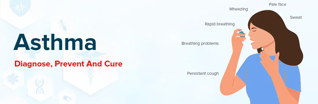
Step 1: Recognizing Symptoms and Taking a Detailed Medical History
The diagnostic journey always begins with a thorough clinical history. Your healthcare provider will ask about current and past respiratory symptoms, their frequency, and their impact on daily life. Key symptoms to discuss include:
-
Wheezing – a high‑pitched whistling sound, most noticeable when breathing out, caused by air passing through narrowed airways.
-
Shortness of breath (dyspnoea) – often worse with physical activity, but can also occur at rest during exacerbations.
-
Chest tightness – a sensation of pressure or constriction in the chest.
-
Cough – typically dry, non‑productive, and often worse at night, early morning, or after exposure to triggers.
Beyond symptom checklists, the provider will explore patterns and variability:
-
Do symptoms come and go, with symptom‑free intervals in between?
-
Are they worse at night or in the early morning (nocturnal or early‑morning dipping)?
-
Are they triggered by specific factors – e.g., viral respiratory infections, exercise, cold air, dust, pollen, pet dander, smoke, strong odours, or emotional stress?
-
Do they improve with bronchodilator medications (e.g., salbutamol)?
A family history of asthma, eczema, hay fever, or other allergic conditions is an important clue, as asthma has a strong genetic component. Also, a personal history of atopic dermatitis or allergic rhinitis increases the likelihood of asthma. The age of onset is relevant: asthma often begins in childhood or early adulthood, whereas new‑onset breathing problems in older adults may suggest COPD or other conditions.
Occupational and environmental exposures should not be overlooked – certain workplaces (e.g., bakeries, chemical plants, farms) can cause or worsen asthma. A detailed medication history, including use of over‑the‑counter cough or cold remedies, is also taken.
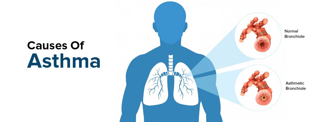
Step 2: Physical Examination
During the physical exam, the healthcare provider will focus on the respiratory system but also look for signs of associated allergic conditions. Steps include:
-
Auscultation – listening to the lungs with a stethoscope while you breathe deeply. The classic finding is expiratory wheezing, but wheezing may be absent when symptoms are well‑controlled or during severe attacks (the “silent chest”).
-
Inspection – observing for signs of increased work of breathing (use of accessory muscles, nasal flaring, tachypnoea) or hyperinflation (barrel chest) in chronic cases.
-
ENT and skin – examining the nasal passages for signs of allergic rhinitis (pale, swollen turbinates) and the skin for eczema or urticaria.
It is important to note that a normal physical examination does not rule out asthma, as many patients have normal findings between episodes. Conversely, wheezing can also occur in other conditions such as COPD, heart failure, or vocal cord dysfunction, so the examination is only one piece of the puzzle.
Step 3: Pulmonary Function Tests – The Cornerstone of Diagnosis
For individuals aged 5 years and older, objective lung function testing is essential to confirm the diagnosis. These tests provide measurable evidence of variable expiratory airflow limitation, which is the hallmark of asthma.
Spirometry
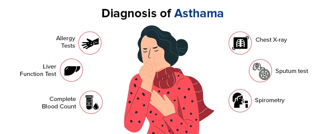
Spirometry is the most widely available and recommended test. You will be asked to take a deep breath and then blow out as hard and as fast as possible into a mouthpiece connected to a spirometer. The key measurements are:
-
Forced Vital Capacity (FVC) – the total volume of air you can forcefully exhale.
-
Forced Expiratory Volume in 1 second (FEV₁) – the volume exhaled in the first second.
-
FEV₁/FVC ratio – this ratio is crucial; a value below the lower limit of normal (usually <0.75–0.80 in adults) indicates obstructive ventilatory impairment.
In asthma, the FEV₁ is typically reduced, and the FEV₁/FVC ratio is low, reflecting airway obstruction. The test is then repeated 15–20 minutes after administering a rapid‑acting bronchodilator (e.g., 400 µg of salbutamol). A significant improvement in FEV₁ (≥12% and ≥200 mL from baseline in adults; ≥12% of predicted in children) is considered a positive bronchodilator reversibility test, which strongly supports the diagnosis of asthma.
However, a normal spirometry result does not exclude asthma – lung function can be normal between exacerbations. In such cases, further testing is needed.
Peak Expiratory Flow (PEF) Monitoring
When spirometry is not available, peak expiratory flow monitoring can be a useful alternative, especially for monitoring variability. You will be given a portable peak flow meter and asked to measure your PEF twice daily (morning and evening) for 1–2 weeks. Record the best of three blows each time. A diurnal variability of >20% (calculated as (highest – lowest)/average × 100) or a clear improvement after bronchodilator is highly suggestive of asthma. PEF monitoring is also valuable for identifying occupational asthma by comparing readings at work and away from work.
Bronchial Challenge Tests (Provocation Tests)
If spirometry is normal and symptoms are intermittent, a bronchial challenge test may be performed to assess airway hyperresponsiveness. The most common method is the methacholine challenge, where you inhale increasing concentrations of methacholine (a substance that causes airway narrowing) and spirometry is repeated. A ≥20% drop in FEV₁ at a low cumulative dose indicates airway hyperresponsiveness, which is typical of asthma but not specific to it (can occur in COPD, allergic rhinitis, etc.). Exercise challenge and cold‑air hyperventilation tests are alternatives. These tests are usually done in specialist centres.
Step 4: Biomarkers and Additional Laboratory Tests
Recent guidelines have emphasised the role of inflammatory biomarkers in supporting the diagnosis and phenotype classification of asthma.
Fractional Exhaled Nitric Oxide (FeNO)
FeNO is a non‑invasive marker of Type 2 airway inflammation. You breathe into a device that measures the concentration of nitric oxide in your exhaled breath. Elevated FeNO levels (≥50 ppb in adults, ≥35 ppb in children) are strongly associated with eosinophilic asthma and can help confirm the diagnosis in patients with suggestive symptoms. However, FeNO can also be raised in non‑asthmatic conditions (e.g., atopy, allergic rhinitis) and may be lowered by smoking or certain medications, so it should be interpreted alongside clinical findings.
Blood Eosinophil Count
A peripheral blood eosinophil count ≥300 cells/µL in adults (or ≥150 cells/µL in some guidelines) supports the diagnosis of Type 2 asthma, especially when combined with other features. This is a simple and widely available test.
Allergy Testing
Allergic sensitisation is a common asthma trait. Skin prick tests or serum‑specific IgE testing for common aeroallergens (house dust mite, pollen, moulds, pet dander) can help identify triggers and guide environmental control. However, a positive test does not prove asthma; it only indicates sensitisation.
Chest Imaging
Chest X‑ray or CT scan is not routinely used to diagnose asthma, but it may be ordered to exclude other conditions such as pneumonia, foreign body aspiration, lung tumours, bronchiectasis, or interstitial lung disease, especially in patients with atypical symptoms or who do not respond to treatment. In chronic severe asthma, a CT may reveal bronchial wall thickening or mucus plugging.
Step 5: Therapeutic Trial (Diagnostic Treatment)
In settings where lung function testing is not feasible or in young children, a diagnostic trial of asthma medication may be appropriate. The patient is given a regular low‑dose inhaled corticosteroid (with or without a long‑acting bronchodilator) for 4–8 weeks, and symptom improvement and/or PEF improvement is monitored. A clear and sustained response supports the diagnosis. However, this approach requires careful follow‑up to avoid over‑ or under‑treatment.
Special Considerations in Specific Populations
Diagnosis in Children Under 5 Years
Young children cannot reliably perform spirometry or FeNO manoeuvres. Therefore, the 2025 and 2026 GINA guidelines recommend a symptom‑based approach combined with a therapeutic trial. The key criteria are:
-
Recurrent episodes of wheezing, cough, or difficulty breathing, especially with viral infections.
-
No alternative explanation (e.g., cystic fibrosis, congenital airway anomalies).
-
Good clinical response to a short course of inhaled bronchodilators and/or corticosteroids.
-
If symptoms persist, referral to a paediatric respiratory specialist is advised.
Diagnosis in Older Adults
In older adults, asthma can be confused with COPD, heart failure, or deconditioning. The presence of a significant bronchodilator response, high FeNO, and a history of atopy favour asthma. However, many older patients have asthma‑COPD overlap (ACO), which requires a tailored diagnostic approach. Always consider cardiovascular causes and drug‑induced cough (e.g., ACE inhibitors).
Diagnosis in Patients Already on Treatment
If you are already taking asthma medications when you are assessed, your lung function may appear normal, and reversibility testing may be negative. In such cases, your doctor may ask you to temporarily withhold certain medications (under supervision) before testing, or they will rely more on biomarkers and historical evidence. The 2026 GINA update provides a separate diagnostic algorithm for patients already on controller therapy.
Differential Diagnosis – Ruling Out Other Conditions
Many respiratory and non‑respiratory conditions can mimic asthma. A thorough evaluation helps to differentiate:
-
COPD – usually later onset, persistent and progressive airflow obstruction, less reversibility, and a strong smoking history.
-
Chronic upper airway cough syndrome (post‑nasal drip) – cough as the main symptom, associated with nasal discharge.
-
Vocal cord dysfunction (inducible laryngeal obstruction) – causes breathlessness and wheeze, but often with inspiratory stridor and no response to bronchodilators; laryngoscopy confirms it.
-
Heart failure – orthopnoea, paroxysmal nocturnal dyspnoea, basal crackles, and cardiomegaly on chest X‑ray.
-
Bronchiectasis – chronic productive cough, clubbing, and characteristic CT findings.
-
Foreign body aspiration – sudden onset of symptoms, especially in children.
-
Pulmonary embolism – acute dyspnoea, pleuritic chest pain, and risk factors for thrombosis.
-
Hyperventilation syndrome – episodic breathlessness, often with paresthesias and panic symptoms.
Your doctor will tailor investigations based on your age, risk factors, and clinical presentation.
Recent Advances and GINA 2025/2026 Updates
The Global Initiative for Asthma (GINA) publishes annual strategy reports that shape clinical practice worldwide. The 2025 update introduced a major change by incorporating Type 2 inflammatory biomarkers (FeNO and blood eosinophils) into the formal diagnostic criteria, which improved diagnostic specificity from 46% to 96% in a large validation study. The 2026 update further simplified the diagnostic flowchart, revised the bronchodilator responsiveness criteria (now using absolute change in FEV₁ alongside percentage change), and provided clear guidance on diagnosing asthma in patients who are already on maintenance therapy. These updates aim to reduce both under‑diagnosis and over‑diagnosis, which remain significant problems globally.
When to See a Healthcare Provider
If you experience recurrent episodes of coughing, wheezing, chest tightness, or breathlessness – particularly if these occur at night, early morning, or after exposure to triggers – you should consult a healthcare professional. Do not ignore persistent symptoms, as early diagnosis allows for timely intervention, reduces the risk of severe exacerbations, and preserves long‑term lung function.
A stepwise, systematic approach combining history, examination, lung function, biomarkers, and when necessary, therapeutic trials, ensures that asthma is diagnosed accurately and that alternative diagnoses are excluded. With the right diagnosis, most people with asthma can achieve good control and live full, active lives.